32 — Work
Neuromuscular orthodontics: what the label can—and cannot—mean
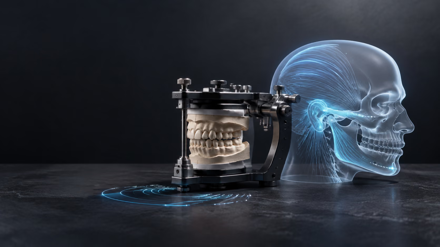
The jaw’s muscles, joints, nerves, teeth, and bite interact, but the label does not define one diagnosis, test sequence, appliance, or treatment.
Original editorial concept · temporary
What it is
“Neuromuscular orthodontics” is a broad label rather than one standardized diagnosis, specialty, test panel, appliance, or treatment philosophy. Teeth, chewing muscles, temporomandibular joints, nerves, and behavior can influence comfort and function, but their interaction does not prove that a bite discrepancy caused pain or that moving teeth will resolve it. A responsible plan separates the orthodontic diagnosis from any TMD or orofacial-pain diagnosis, records symptoms before movement, uses imaging and instrumented measurements only for defined questions, favors conservative and reversible TMD care when appropriate, and recommends orthodontic treatment only for a supported orthodontic objective.
For
01
A person considering orthodontic tooth movement while experiencing jaw pain, locking, limited motion, painful joint sounds, headache, or a changing bite.
02
Someone asked to interpret computerized jaw tracking, surface electromyography (sEMG), transcutaneous electrical nerve stimulation (TENS), a bite record, or an appliance as part of an orthodontic or TMD evaluation.
03
Anyone evaluating a claim that changing tooth position or the bite will prevent or cure a temporomandibular disorder, headache, sleep-disordered breathing, an airway condition, or posture symptoms.
01 — Clarify the question before selecting a label
The first task is to identify what the patient wants assessed: tooth position, bite, jaw pain, joint noise, locking, limited motion, headache, sleep symptoms, restorative planning, or another concern. Each question has its own history, examination, evidence, and referral boundary; the word neuromuscular cannot answer them together.
02 — Record symptoms and function before movement
Jaw pain, headache, joint sounds, locking, range and pattern of motion, chewing limits, parafunctional habits, trauma, sleep and medical history, previous appliances, and current medicines should be documented before orthodontic treatment. A baseline helps distinguish a pre-existing or fluctuating condition from a symptom that appears or changes during care.
03 — Diagnose the orthodontic and TMD questions separately
Orthodontic assessment addresses teeth, eruption, growth, skeletal relationships, supporting tissues, movement, and retention. TMD assessment addresses pain and joint or muscle disorders through history and clinical examination, with validated criteria and appropriate differential diagnosis. One diagnosis should not be inferred from the other.
04 — Select imaging and adjuncts for a defined decision
Existing records are reviewed first. Radiographs, CBCT, MRI, jaw tracking, surface EMG, or another measure should be selected only when the expected information addresses a stated question and its limitations are explained. A tracing, activity graph, or device-generated position is not a diagnosis by itself.
05 — Treat conservatively and coordinate when pain is present
Education, self-management, appropriate physical therapy, medicines prescribed for the individual, and selected reversible appliances may be considered for TMD according to the diagnosis. Persistent, complex, neurologic, systemic, traumatic, or atypical findings can require referral to an orofacial-pain clinician, physician, physical therapist, surgeon, or another qualified professional.
06 — Move teeth for an orthodontic objective, then monitor and retain
If orthodontic treatment is appropriate, the plan should name the intended tooth or jaw relationship, alternatives, biological limits, risks, monitoring, and retention. New or changing pain, locking, motion, bite, tooth, periodontal, or neurologic findings require reassessment rather than an assumption that the original explanation remains correct.
Orthodontics and TMD guide
A responsible plan separates symptoms, diagnosis, measurements, orthodontic objectives, and TMD care.
The useful consultation is not a search for one machine-generated jaw position. It explains which problem is being evaluated, which findings support each diagnosis, what uncertainty remains, whether a conservative trial is appropriate, how tooth movement would serve an orthodontic objective, and when another clinician should participate.
“Neuromuscular” is descriptive language—not one diagnosis
The term can refer broadly to the relationship among nerves, muscles, joints, teeth, and movement. Different clinicians and commercial systems may use it differently. It does not identify a validated disease entity, a universal examination, a necessary sequence of devices, a single appliance design, or an assured treatment result.
The label is not a separately recognized dental specialty
The American Dental Association’s current recognized dental specialties include Orthodontics and Dentofacial Orthopedics and Orofacial Pain; neuromuscular dentistry is not listed as a separate specialty. Training, licensure, scope, and referral responsibility therefore must be stated accurately for the clinician involved rather than inferred from the label.
Orthodontic diagnosis and TMD diagnosis answer different questions
An orthodontic diagnosis describes tooth position, eruption, growth, skeletal relationships, supporting tissues, and the movement that may be appropriate. A TMD diagnosis distinguishes muscle pain, joint pain, disc-related conditions, degenerative change, headache attributed to TMD, and important mimics. A person can have either, both, or neither.
Baseline symptoms and function belong in the record
Jaw pain, temple headache, joint sounds, locking, limited opening, deviation, chewing difficulty, clenching, grinding, trauma, sleep, stress, previous treatment, medical conditions, and medicines should be recorded before tooth movement. Symptoms often fluctuate, so timing, frequency, intensity, triggers, limitations, and prior episodes are more informative than a single yes-or-no answer.
History and examination carry the diagnosis
Current professional guidance places the patient history and clinical examination at the center of TMD assessment. Palpation, movement, pain provocation, joint sounds, oral and dental findings, cranial-nerve or other screening, and differential diagnosis are selected for the presentation. A device printout cannot replace those findings or exclude dental, neurologic, rheumatologic, infectious, traumatic, vascular, or other causes.
Validated criteria structure assessment; they do not automate it
The Diagnostic Criteria for Temporomandibular Disorders (DC/TMD) provide validated symptom and examination algorithms for common pain-related and intra-articular TMDs together with assessment of pain-related disability and psychosocial factors. They support consistent reasoning; they do not make every case simple, eliminate clinical judgment, or establish an orthodontic indication.
The bite is not the default cause of jaw pain
Current evidence does not support treating ordinary malocclusion or one occlusal feature as a strong universal cause of TMD. Associations reported in observational studies can reflect confounding, reverse causation, selection, or measurement differences. A changed contact may matter in an individual examination, but it does not establish that irreversible bite alteration will treat the disorder.
Orthodontics is not a general TMD treatment
Systematic reviews have not established that orthodontic treatment prevents TMD or reliably treats it as a category. Tooth movement can be appropriate for a diagnosed orthodontic problem in a person who also has TMD, but it should not be presented as a cure for jaw pain, headache, ear symptoms, sleep-disordered breathing, airway disease, posture, or systemic symptoms.
Association is not causation—and the literature contains disagreement
A 2022 meta-analysis reported an association between orthodontic treatment and TMD, while a published critique challenged its methods and interpretation; earlier systematic reviews found no clear cause-and-effect relationship. The disagreement does not justify claiming that orthodontics causes, prevents, or cures TMD. It supports documenting baseline findings, explaining uncertainty, monitoring change, and reassessing symptoms.
Jaw tracking records movement, not a diagnosis
Computerized mandibular tracking can describe a recorded movement path under defined conditions. Systematic reviews report limited diagnostic reliability, small or heterogeneous studies, risk of bias, and insufficient validation against accepted reference standards. A tracing may supplement a question, but it cannot establish disease, causation, an ideal jaw position, or a required treatment by itself.
Surface EMG records electrical activity, not a diagnosis
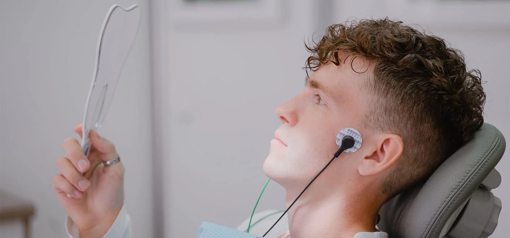
Surface electromyography records electrical activity from selected muscles through skin electrodes. Electrode placement, anatomy, skin conditions, tasks, effort, pain, fatigue, signal processing, and reference values can change a recording. Reviews have not established adequate standalone diagnostic performance for TMD, so sEMG should not be described as locating a uniquely correct bite or proving treatment need.
TENS is a treatment adjunct, not a diagnostic test
Transcutaneous electrical nerve stimulation applies electrical stimulation through surface electrodes and may be considered as a pain-modulation adjunct for selected patients. Evidence for TMD is heterogeneous and does not support using a response to TENS to diagnose the disorder, reset the muscles, reveal a permanent ideal jaw position, guarantee relief, or determine irreversible orthodontic or restorative treatment.
Imaging is selected for a defined question
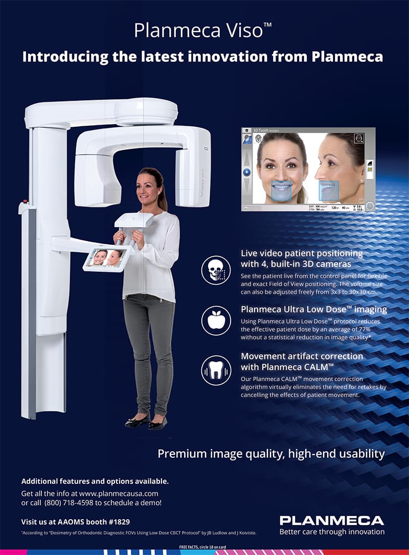
Many TMD assessments do not begin with advanced imaging. Imaging is considered when history and examination indicate that information about bone, joint structure, disc position, inflammation, trauma, growth, or another feature may change diagnosis or management. Existing images and radiation exposure should be reviewed before ordering another study.
MRI and CBCT answer different joint questions
MRI is generally the preferred modality for articular-disc position and other soft-tissue or inflammatory questions. CBCT or CT is more suitable for osseous morphology and degenerative bone change. Neither is a universal screening test, and imaging findings can occur without matching symptoms; interpretation must remain connected to the clinical question.
An appliance trial does not establish an ideal jaw position
A reversible appliance may be used for selected protection, symptom management, or diagnostic observation, but improvement or change during a trial does not prove one permanent condylar or occlusal position. Appliances can also alter comfort, contacts, function, or symptoms and require monitoring. A reversible response should not be converted automatically into tooth movement, equilibration, crowns, or surgery.
Conservative and reversible TMD care comes first when appropriate
Professional guidance favors evidence-based, conservative, reversible approaches for many TMDs. Depending on the diagnosis, this can include explanation, self-management, behavior and habit support, appropriate physical therapy, and individualized medicines or appliances. Exact recommendations, contraindications, and escalation depend on the person; persistent or complex disease may require specialist or medical care.
Orthodontic objectives still require an orthodontic diagnosis
Crowding, spacing, eruption, crossbite, overbite, overjet, missing-tooth space, skeletal discrepancy, and restorative preparation can create legitimate orthodontic questions. The plan should name the intended movement, expected benefit, accepted compromise, anchorage, appliances, alternatives, and what will not be changed. A TMD label or device measurement is not a substitute for that diagnosis.
Growth, supporting tissues, roots, and restorations set boundaries
Age and growth, periodontal inflammation and bone support, caries, enamel, root form or resorption, previous trauma, implants, crowns, bridges, missing teeth, and planned restorative care can change candidacy, force, sequence, monitoring, and prognosis. Healthy or stabilized tissues and coordinated restorative objectives come before irreversible movement.
New or changing symptoms require reassessment
Pain, locking, limited opening, joint sounds, bite change, tooth mobility, numbness, weakness, swelling, fever, trauma, or a new neurologic symptom should not be dismissed as a normal adjustment without assessment. The treating team should decide whether to pause or modify movement, obtain records, manage a dental cause, or refer for TMD, medical, neurologic, or surgical evaluation.
Referral can be the correct next step
Orofacial-pain clinicians, orthodontists, general dentists, oral and maxillofacial surgeons, physical therapists, physicians, radiologists, neurologists, rheumatologists, sleep clinicians, and behavioral-health professionals answer different questions. Referral is appropriate when the diagnosis, risk, or treatment lies beyond the responsible clinician’s competence or when coordinated care would improve safety and clarity.
Risks, alternatives, and retention remain part of orthodontic care
Possible orthodontic burdens include soreness, soft-tissue irritation, enamel demineralization or caries, gingival inflammation, recession or attachment loss in susceptible tissues, root resorption, pulp change after trauma, unwanted movement or bite change, prolonged or incomplete treatment, relapse, and additional procedures. Alternatives can include observation, limited treatment, fixed or removable appliances, restorative care, surgery or referral, and no active movement. Retention and follow-up remain necessary after active care.
Emergency features change the response
Difficulty breathing or swallowing, choking or suspected aspiration, collapse, confusion, new facial weakness, sudden severe neurologic symptoms, rapidly progressive facial or neck swelling, heavy uncontrolled bleeding, or major facial trauma requires 911 or the nearest emergency department. Fever with worsening swelling, suspected jaw fracture, a knocked-out or severely displaced permanent tooth, or an acutely locked jaw requires prompt professional assessment. A website or remote monitor is not an emergency service.
Equipment, training, and local availability must be verified
This educational page does not establish that TetriSmile offers a neuromuscular service, uses K7 or another jaw-tracking, sEMG, TENS, imaging, or appliance system, or has a clinician with a particular specialty or credential. Exact equipment, software, calibration, training, protocol, interpretation, decision impact, fees, timing, outcomes, and lawful service scope require current signed practice records before publication or scheduling claims.
Continue the evaluation
Clinical reading
General information only. Diagnosis, treatment, risks, alternatives, and expected recovery are individual and require a clinical evaluation.
- NIDCR: Temporomandibular Disorders ↗
- NIDCR: Summary of treatments for TMDs ↗
- AADOCR/IADR: Temporomandibular disorders position statement ↗
- Diagnostic Criteria for Temporomandibular Disorders (DC/TMD) ↗
- AAOMR/AAOP: Imaging of the temporomandibular joint ↗
- National Academies: Temporomandibular Disorders ↗
- British Orthodontic Society: TMD and the orthodontic patient ↗
- ADA: Recognized dental specialties ↗
- Systematic review: Orthodontic treatment and TMD ↗
- Systematic review: Occlusion and TMD ↗
- Systematic review: Baseline TMD findings in orthodontic patients ↗
- Meta-analysis: Orthodontic treatment and TMD association ↗
- Published critique of the orthodontic-TMD meta-analysis ↗
- Systematic review: Surface EMG in TMD diagnosis ↗
- JADA systematic review: Surface EMG diagnostic efficacy ↗
- Systematic review: Computerized jaw tracking ↗
- Systematic review: Jaw tracking in TMD ↗
- Systematic review: TENS for temporomandibular disorders ↗
- Controlled evidence: TENS and TMD pain ↗
A responsible discussion should make nine things clear: the patient’s question, separate orthodontic and TMD diagnoses, baseline symptoms, the limits of imaging and instrumented measurements, reversible-first care when appropriate, the reason for any tooth movement, referral boundaries, realistic risks and alternatives, and the retention and reassessment plan.